Promotion
Use code SIZZLE26 for 25% off sitewide!

By clicking “Accept,” you agree to the use of cookies and similar technologies on your device as set forth in our Cookie Policy and our Privacy Policy. Please note that certain cookies are essential for this website to function properly and do not require user consent to be deployed.
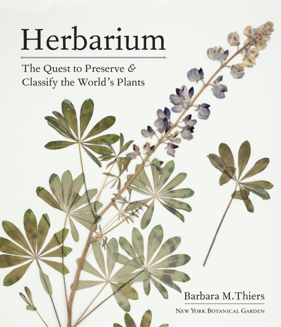
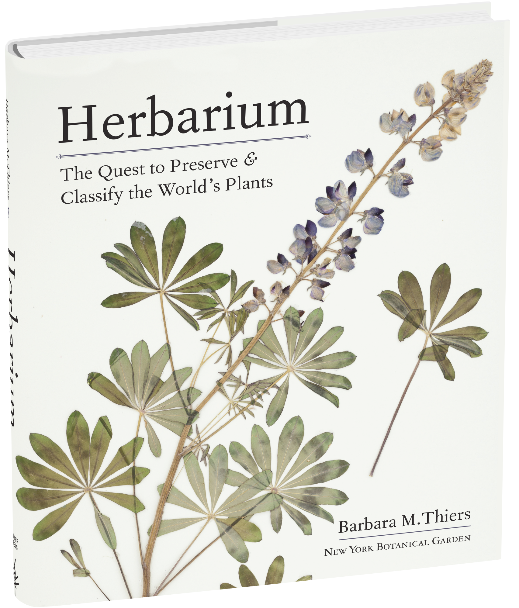
Herbarium
The Quest to Preserve and Classify the World's Plants


Contributors
Formats and Prices
- On Sale
- Dec 8, 2020
- Page Count
- 304 pages
- Publisher
- Timber Press
- ISBN-13
- 9781604699302
Price
$40.00Price
$50.00 CADFormat
Format:
- Hardcover $40.00 $50.00 CAD
- ebook $18.99 $24.99 CAD
This item is a preorder. Your payment method will be charged immediately, and the product is expected to ship on or around December 8, 2020. This date is subject to change due to shipping delays beyond our control.
Buy from Other Retailers:
“A sweeping history of the origins, development, and future of herbaria and their role in plant consternation.” —The American Gardener
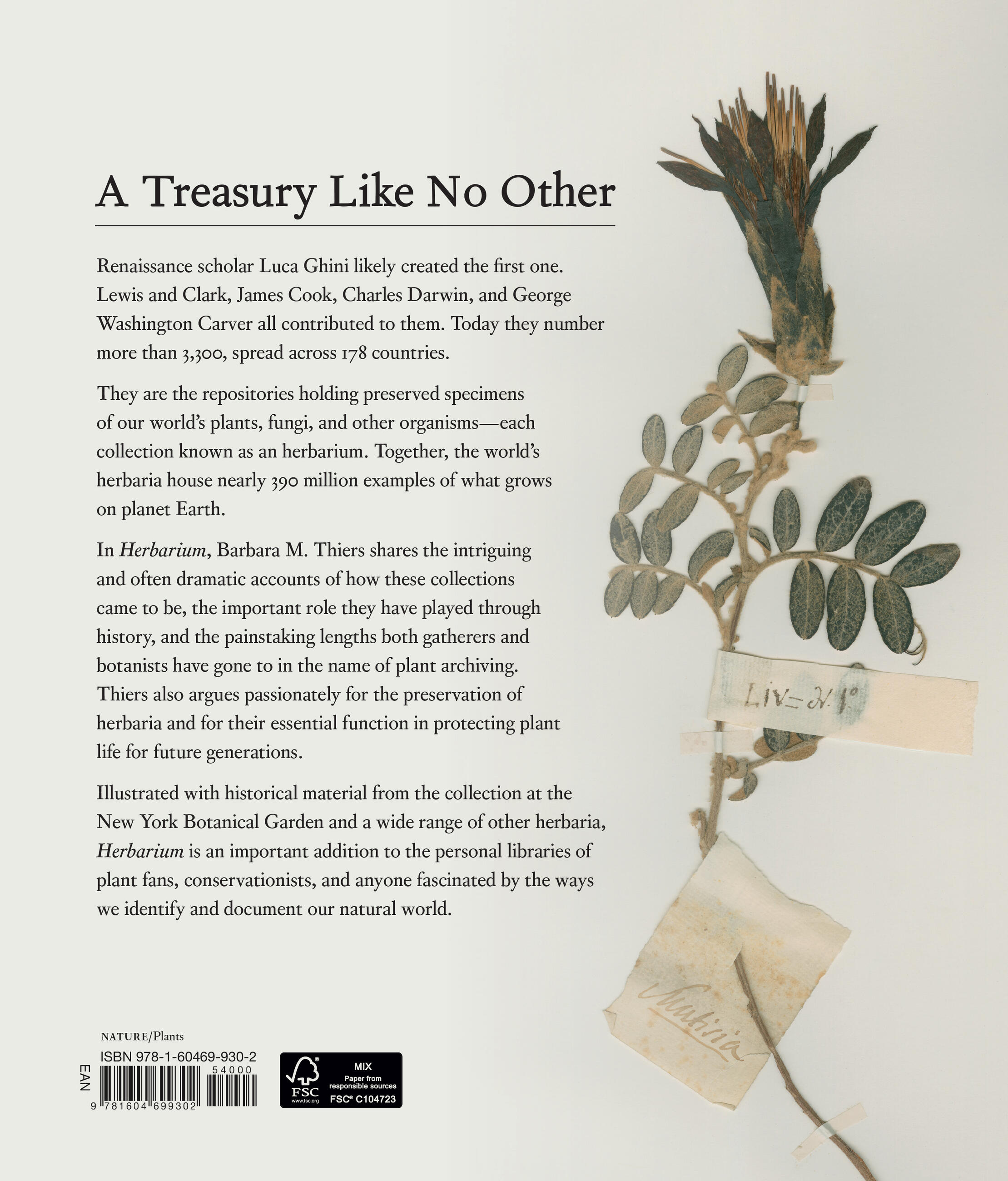
Since the 1500s, scientists have documented the plants and fungi that grew around them, organizing the specimens into collections. Known as herbaria, these archives helped give rise to botany as its own scientific endeavor.
Herbarium is a fascinating enquiry into this unique field of plant biology, exploring how herbaria emerged and have changed over time, who promoted and contributed to them, and why they remain such an important source of data for their new role: understanding how the world’s flora is changing. Barbara Thiers, director of the William and Lynda Steere Herbarium at the New York Botanical Garden, also explains how recent innovations that allow us to see things at both the molecular level and on a global scale can be applied to herbaria specimens, helping us address some of the most critical problems facing the world today.
Genre:
-
“With lavish illustrations of places and people; portraits of key players; herbaria specimens; and beautiful, full-color artists’ renderings, this carefully researched, detailed homage to herbaria will appeal to those deeply interested in plant exploration and botany.” —Library JournalThe Botanical Society of Britain and Ireland
“A sweeping history of the origins, development, and future of herbaria and their role in plant consternation.” —The American Gardener
“One of the prettiest books of the season… a lovely coffee table book as well as a serious work on the history of scientific endeavor.” —The Napa Valley Register
“An enlightening tribute to the mysterious world of Herbaria that is meticulously researched, well organized, and above all accessible.” —Gardens Illustrated
“This illustrated account of the Herculean efforts of the early botanists in this field is absorbing and enlightening.” —The English Garden
“At its heart, Herbarium is a compelling reminder of one of humanity’s better impulses: to save things—not just for ourselves, but for posterity… A marvelous book about the ages, for the ages.” —The Nevada Native Plant Society
“Herbarium is sure to enchant anyone who has ever used a flower press or wished to learn more about plant classification, with vintage engravings and woodcuts of ancient plants.” /B>The Free Press
“Invaluable for any personal library on the history of plants and their future. It is beautifully produced.” —The Financial Times
“Gardeners with an interest in history will delight in Herbarium.” —Horticulture
“Thoroughly researched and written in a readable style…this coffee-table book is lavishly illustrated with herbarium specimens, people, places, and maps, it’s one of those glorious books where hours can easily be lost in reading and re-reading.” —RHS The Garden
“A great book on the history of herbaria…readers interested in biographies and history would find this a great book to read and to add to their library collection.” —Cultivate to Plate
“This beautifully illustrated history includes not just full-page examples of herbaria, but paintings, prints, maps, and photographs.” —Landscape Architecture
“This book gives life to botanical collections and is a brilliant work that can be read, understood, and enjoyed by everyone.” —The Journal of the American Botanical Council
“A journey through the history of botanical collecting… those vivid stories bring specimens to life.”—Plant Science Bulletin
“This sumptuously illustrated book will delight all botanists, whether their hearts lie in the field or in the herbarium…This is a book that belongs on every botanist’s bookshelf, for the wonderful stories contained within and for the marvelous illustrations of specimens that grace its pages.”