Promotion
Use code DAD23 for 20% off + Free shipping on $45+ Shop Now!
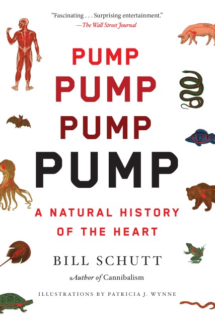
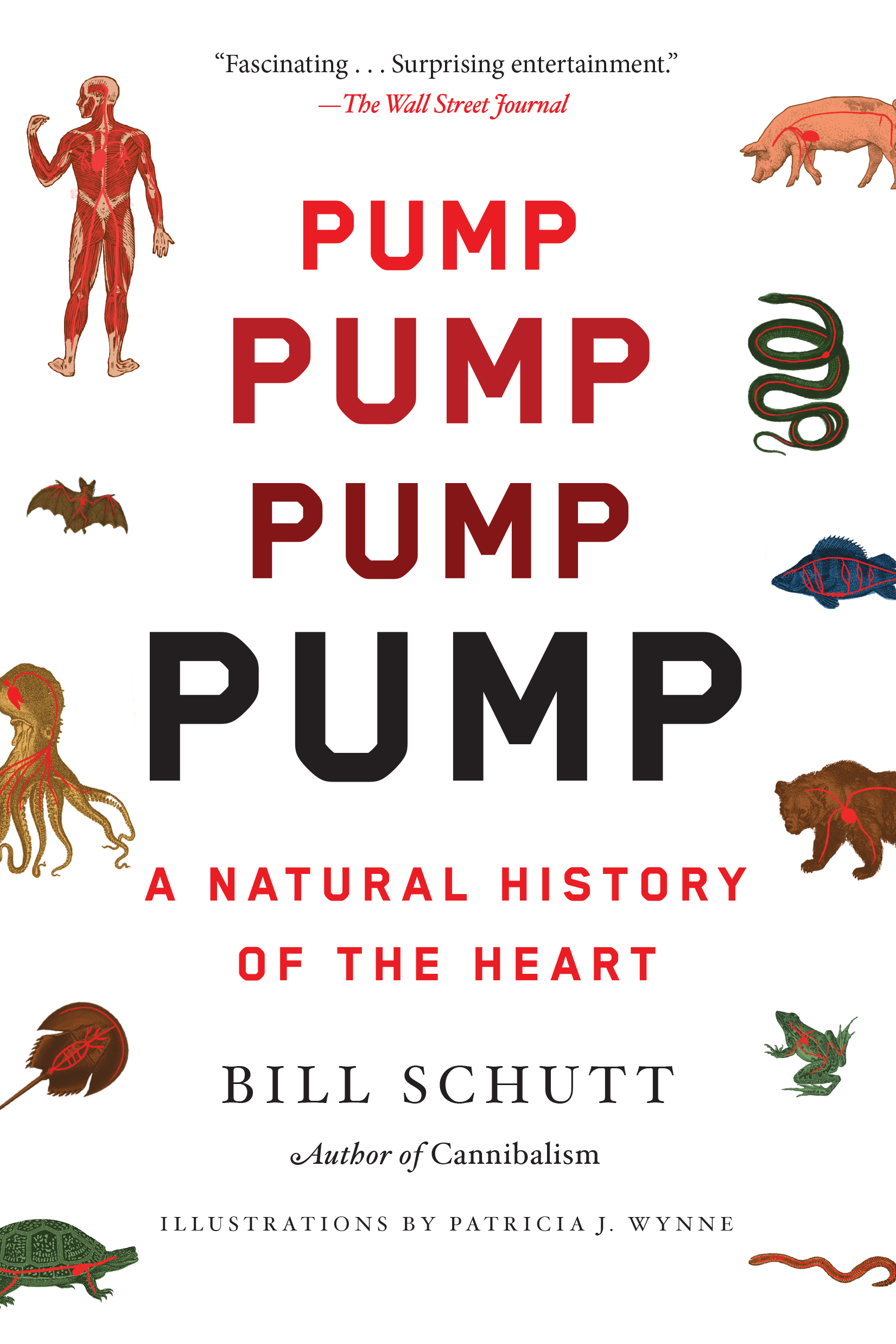
Pump
A Natural History of the Heart
Contributors
By Bill Schutt
Formats and Prices
Price
$17.99Price
$22.99 CADFormat
Format:
- Trade Paperback $17.99 $22.99 CAD
- ebook $11.99 $15.99 CAD
- Audiobook Download (Unabridged)
This item is a preorder. Your payment method will be charged immediately, and the product is expected to ship on or around September 13, 2022. This date is subject to change due to shipping delays beyond our control.
Also available from:
"Fascinating . . . Surprising entertainment, combining deep learning with dad jokes . . . [Schutt] is a natural teacher with an easy way with metaphor.”—The Wall Street Journal
In this lively, unexpected look at the hearts of animals—from fish to bats to humans—American Museum of Natural History zoologist Bill Schutt tells an incredible story of evolution and scientific progress.
We join Schutt on a tour from the origins of circulation, still evident in microorganisms today, to the tiny hardworking pumps of worms, to the golf-cart-size hearts of blue whales. We visit beaches where horseshoe crabs are being harvested for their blood, which has properties that can protect humans from deadly illnesses. We learn that when temperatures plummet, some frog hearts can freeze solid for weeks, resuming their beat only after a spring thaw. And we journey with Schutt through human history, too, as philosophers and scientists hypothesize, often wrongly, about what makes our ticker tick. Schutt traces humanity’s cardiac fascination from the ancient Greeks and Egyptians, who believed that the heart contains the soul, all the way up to modern-day laboratories, where scientists use animal hearts and even plants as the basis for many of today’s cutting-edge therapies.
Written with verve and authority, weaving evolutionary perspectives with cultural history, Pump shows us this mysterious organ in a completely new light.
Excerpt
HEART (n.)
1 A hollow muscular organ that pumps the blood through the circulatory system by rhythmic contraction and dilation. In vertebrates, there may be up to four chambers (as in humans), with two atria and two ventricles.
2 Used to refer to a person's character, or the place within a person where feelings or emotions are considered to come from.
3 The firm central part of a vegetable, especially one with a lot of leaves.
4 Courage, determination, or hope.
5 A shape consisting of two half circles next to each other at the top and a V shape at the bottom, often colored pink or red and used to represent love.
6 One of the four suits in playing cards, represented by a red heart shape.
7 The central or most important part.
Part 1
Wild at Heart
One size does not fit all.
—unknown (possibly Frank Zappa)
[ 1 ]
Size Matters I
In august 2018, I traveled to Toronto's Royal Ontario Museum with artist Patricia J. Wynne to examine the famous blue whale heart. Patricia and I have been friends and officemates at the American Museum of Natural History since the mid-1990s, and she has illustrated every paper, book chapter, and book (fiction and nonfiction) that I've ever written. Although the blue whale exhibit had already closed and the specimen was being stored at an off-site facility, researcher Bill Hodgkinson had uncrated the heart in preparation for our arrival. In a room the size of a small aircraft hangar, the preserved whale heart sat perched upon a two-inch-thick stainless-steel rod, giving it the appearance of having been skewered from below. The bottom end of the skewer was secured to a wooden floor stand while the business end had been connected to a metal armature, invisible to viewers, that served as the heart's permanent internal scaffold.
Because the specimen's official dimensions are forty-two inches from top to bottom by thirty-eight inches in width, I was quite surprised to find it looming over me at a height of what I estimated to be well over six feet. The explanation for the added height was the massive blood vessels situated atop the plastinated organ. The most prominent of these was the great arch of the aorta and its offshoots, a pair of carotid arteries that had once carried oxygenated blood from the left ventricle of the heart to the animal's head. If the previously mentioned atria can be envisioned as the heart's receiving chambers (the left atrium and the right atrium receiving blood from the lungs and the body, respectively), then the ventricles are the heart's pumping chambers—the right ventricle pumps oxygen-poor/CO2-rich blood to the lungs while the left pumps oxygenated blood out to supply the body.
During the blue whale heart's lengthy preparation period, a special type of colored silicone polymer had been injected into the blood vessels, and so veins and arteries could be now be differentiated, because veins were blue and arteries were red. The multicolored heart was really quite beautiful, and I was immediately drawn to a porthole-shaped section that had been cut through the right ventricle by plastination expert Vladimir Chereminsky. The window allows viewers to peer inside the chamber, where, among other things, they can see the odd-looking arrangement of inch-thick muscle strands that line its walls. These strands are known as trabeculae carneae (meaty ridges) by anatomy types and medical professionals, and smaller versions can be seen in many mammals, including humans. The ridges increase the surface area of the ventricular walls as compared to a smooth wall, packing more muscle fibers into a limited space. This is important because the extra muscle translates to stronger ventricular contractions, which propel blood out of the heart. Additional functions of this odd-looking chamber surface remain to be explored.
The right and left atria of the whale heart also contract, and their thinner walls reflect the fact that their job is less difficult: pumping blood into their adjacent ventricles instead of out to the body. Located between the atria and ventricles are the aptly named atrioventricular (AV) valves. Through Chereminsky's porthole, museum visitors could see the blue whale's right AV valve, which appeared to have the diameter of a toddler's toy drum. In humans, the corresponding valve spans about three-quarters of a square inch, about the diameter of a marble, and is more commonly known as the tricuspid valve, due to its three flap-like valve cusps.*
The AV valves regulate blood flow from the atria to the ventricles, but equally important is their job preventing blood from reversing direction and heading back into the atria when the ventricles contract. Vital to this role, and clearly visible within the blue whale heart, are a dozen or so tough fibers known as chordae tendineae. Colloquially known as the heartstrings (since they resemble pieces of string), these cords are composed primarily of a structural protein called collagen.† With one end of the chordae tendineae firmly anchored to the floor of the ventricle and the other end attached to the valve cusp, the cusps are prevented from extending into the atria when the ventricles contract—effectively sealing off the two chambers.
To visualize this, picture a dog with its collar fastened to a long leash, with the nondog end staked to the ground. The dog (standing in for the valve cusps) can travel only so far before the leash (the chordae tendineae) pulls tight, preventing the dog from advancing past an open gate. In humans, the terms "ventricular prolapse" or "prolapsed valve" are used to describe medical conditions in which one or more of the AV valve cusps bulges into an atrium (think of the dog's leash that has been stretched from the pup's constant tugging, allowing it to advance beyond the gate). Since this prolapse breaks the seal separating the atrium and the ventricle, some of the ventricular blood "regurgitates" back into the atrium when the ventricle contracts, instead of leaving the heart, as it would normally. These so-called "floppy" valves can result from previous heart attacks, infections like bacterial endocarditis (frequently found in intravenous drug users), or rheumatic fever, a now-rare consequence of untreated strep throat or scarlet fever. Mitral valve prolapse can also be congenital in nature.
Valve problems can also be a consequence of aging. As the heart valves stiffen and lose their flexibility, they lose their ability to efficiently seal off the heart chambers. With some of the blood moving backward into the atrium with each heartbeat, less blood is pumped out of the heart, and so it has to work harder (by increasing its rate or contracting harder) to compensate. The extra effort can put added stress on the heart, which can lead to serious problems. These become especially apparent if the heart reaches a point at which it can no longer provide sufficient oxygen- and nutrient-rich blood to the body.
Once blood passes through the AV valves, filling the right and left ventricles, it must next pass through the semilunar valves, named for their half-moon-shaped cusps. As the ventricles contract, blood rushes through them into two large arteries. On the right side is the pulmonary trunk, which sends deoxygenated blood to the lungs via the pulmonary arteries that branch from it. On the left side, ventricular contraction pumps oxygenated blood out through the aorta, whose branches distribute it to the rest of the body. Though their anatomy is different from the AV valves before them—no chordae tendineae here—pulmonary and aortic semilunar valves also prevent the backflow of blood, here from the pulmonary artery and aorta back into the ventricles.
In humans, slight valvular abnormalities are often symptom-free and don't require treatment. In more serious cases, a prolapsed valve can cause irregular heartbeat (arrhythmia), dizziness, fatigue, and shortness of breath, and surgery may be required to fix it. Until the early 2000s, valve repair or replacement required complicated open-heart surgery. Now, though, transcatheter valve replacements can be accomplished through small incisions, or even no incisions at all, as a result of major advances in cardiac catheterization—a process whose history is as interesting as any fiction writer could have dreamed up. But more on that topic later.
To give viewers a look at the blue whale's heart just below its surface, plastination-meister Chereminsky also removed a section of the whale's visceral pericardium. This is the thin, protective layer of the heart that lies atop all that muscle. It's also the inner layer of the saclike pericardium, which lubricates and cushions the heart while holding it in place. To visualize the relationship between heart and the pericardium, picture a Ziploc storage bag containing a bit of water. Push your fist (the heart) into the side of the bag so that the bag wraps around your fist. The bag of water is the pericardium, and the part of the bag plastered against your fist is the visceral pericardium. The space inside the bag is the pericardial cavity, partially filled with its supply of pericardial fluid. To complete the metaphor, the part of the Ziploc bag farthest from your fist is the parietal pericardium, and it is attached to the surrounding walls of the chest cavity. This connection anchors the heart in place while cushioning it from external shocks. It's worth noting that the pericardium does not contain the heart, but rather is wrapped around it.
Having observed the plastinated whale heart, inside and out, I left my friend Patricia at the warehouse to sketch the specimen while I set off for the ROM to interview some of the people responsible for its recovery and preservation. But beyond the story of how this one-of-a-kind specimen came to be, I was most interested in what Jacqueline Miller, Mark Engstrom, and their colleagues learned from it that they had not known before.
I asked Miller about the plastinated heart's odd shape. Typically, the mammalian heart is conical, coming to a single point at the bottom or apex. I had been struck by the fact that in blue whales the apex of the heart is split. Miller explained that this bifurcation is a characteristic of rorquals, a name used to group the largest of the baleen whales.* Another unique characteristic, she told me, is that this particular heart is flatter and wider than most mammalian hearts.
"The typical terrestrial mammal has a spiral heart—a heart in which the connective tissue and muscle fibers are oriented so that they spiral around the left and right ventricles," added Engstrom. "When the heart contracts, the overall action is more like wringing out a towel."
But in rorquals, the fibers run straight from the top of the heart (the base) to the bottom, rather than in a spiral.
"I think what's happening is that when they do deep dives, their heart collapses," Engstrom told me.† "It's still beating, but it collapses due to the pressure."
Because of this, and as Miller and her team discovered back in Rocky Harbour, once the heart had been severed from its moorings and removed from the body, it had collapsed "like an enormous spongy bag," according to Miller, thus requiring reinflation during the preservation process.
Adding to the list of things the researchers at the ROM had learned about blue whales, Engstrom mentioned how many times over his career he had been asked about the actual size of the world's largest heart.
"I was getting tired of the question," he admitted. "And I really wanted to be able to say 'It's that big' and then point to it."
For decades, in both popular and scientific literature, it was written that a blue whale heart would be the size of a sedan and weigh at least a metric ton.‡ Miller told me that in preparing to extract the heart, she and her colleagues had read about how "you'd be able to swim down one of the greater vessels, presumably the caudal vena cava, which is the largest vessel on the blue whale heart."
As I looked over the impressive vasculature attached to the ROM specimen, it was easy to see that even the largest blood vessel wasn't wide enough for a human to swim through, though I figured an otter or a migrating salmon could make the journey with relative ease.
Indeed, Miller told me, once the heart had been preserved it was significantly smaller than they had thought it would be. And this wasn't an undersized blue whale by any stretch. So why was it so much smaller than anticipated?
The answer turned out to be that blue whale hearts are simply not as large as the hearts of most other mammals. While quite humongous by human standards, a blue whale's heart apparently makes up only around 0.3 percent of the animal's total body weight. For comparative purposes, the relative size of the heart in both mice and elephants has been calculated to be about 0.6 percent.
Interestingly, some of the world's smallest animals have disproportionately large hearts. For example, the masked shrew (Sorex cinereus) is one of the smallest mammals in the world, weighing in at around five grams,* but its heart makes up about 1.7 percent of its body weight, which is approximately three times larger than one would predict for a typical terrestrial mammal, and nearly six times the relative size of a blue whale heart. Birds, meanwhile, tend to have relatively larger hearts than mammalian hearts, due to the metabolic demands of flight. In hummingbirds, the smallest of which can weigh as little as two grams (less than a dime), the heart-to-body weight numbers are even more extreme, with the heart reaching 2.4 percent of body weight. Relatively speaking, this means that hummingbird hearts are eight times larger than those of a blue whale.
It is thought that the reason for possessing a relatively large heart relates to the lifestyles of the small and hyperactive. For example, hummingbirds can beat their wings at eighty times per second, and shrews are such nonstop hunters that during my mammal-trapping days as a PhD student at Cornell University I was taught that they would starve to death if not removed from a live trap within an hour. The manic behavior of these tiny animals causes an extremely high cellular demand for both energy and oxygen. These metabolic requirements are met in part by increasing heart rate, thus also increasing the frequency at which oxygen-rich and nutrient-laden blood is pumped to the body. The resulting heart rate numbers are truly astonishing. Hummingbird heart rates can reach 1,260 beats per minute, while shrews hold the vertebrate record at 1,320 beats per minute—roughly seven times the maximum heart rate of a thirty-five-year-old human.
Though these are eye-popping numbers, the increase in beat frequency is not unlimited, and researchers believe that there is a maximum rate at which a heart can beat. For a shrew, one heartbeat lasts forty-three milliseconds—that's forty-three thousandths of a second. During this split second, the heart needs to fill with venous blood, contract and eject the arterial blood, and relax in preparation for the next filling cycle. All of that can occur only so fast—and if shrews aren't at the upper limit of heart rate, then they're awful damn close. So if the physical design of the heart limits it to something like a maximum of fourteen hundred beats per minute, then the only way to pump more blood is to increase the size of the heart. That way, the larger chambers are able to receive and pump a relatively greater measure of blood with each beat.* This explains the comparatively enormous heart size of creatures like shrews and hummingbirds. But as we'll soon see, increasing heart size among the ubersmall also has its limits.
Before leaving blue whale hearts, though, and whale hearts in general, it should be emphasized that there is much, much more to learn: How exactly do these hearts collapse, and how can their owners survive when they do? Other diving mammals, like seals, reduce their heart rates and cut off blood flow to different regions of their body. Do blue whales possess the same oxygen-saving adaptations? Initial research indicates that this could be so, since a recent study by biologist Jeremy Goldbogen and his colleagues at Stanford University found that blue whale heart rates can drop to as low as two beats per minute.* On the anatomy side of things, other serious questions remain, some as simple as identifying the blood vessels in the confusing assemblage sprouting from the now-famous ROM specimen. Until more research can be done, much of the physiology of the rorqual heart will remain in the realm of hypothesis and conjecture.
* On the left side, the bicuspid valve is named for its two cusps. Confusing the issue, it is also known as the mitral valve, due to its supposed resemblance to a miter, the ceremonial headwear worn by bishops. Thankfully, there are no hat-derived alternative names for the tricuspid valve.
† Wound into fibers, collagen is the most abundant protein in mammals. It is commonly found in tendons, ligaments, and the skin. Collagen also give bones their varying degrees of flexibility.
* "Baleen" is the arrangement of bristles inside the mouths of certain whale species into a filter-feeding device. Composed of keratin (the stuff that makes up our nails and hair), it is used to trap krill after large gulps of water are taken in and then forced out of the mouth.
† While the dive record for a tagged blue whale is 315 meters (1,033.5 feet), a Cuvier's beaked whale (Ziphius cavirostris) holds the record for dive depth by a mammal, at 2,992 meters (or 1.86 miles)!
‡ 1 metric ton = 2,204.6 pounds.
* The smallest mammal in the world is the Kitti's hog-nosed bat (Craseonycteris thonglongyai) from Thailand and Myanmar. Also known as the bumblebee bat, it weighs in at barely two grams.
* An average-sized man has about five liters of blood. At rest, cardiac output is approximately five liters/minute, so the average time it takes our blood to take a full circuit of the body (from heart to lungs, back to the heart, out to the body, and back to the heart) is approximately one minute.
* Goldbogen and his team used suction cups to attach a heart rate monitor to a single blue whale, and were able to monitor the animal's heart rate for nearly nine hours. They did not seek to determine if blood flow was redirected to specific regions of the body during the dramatic drop in heart rate that they recorded.
The Microbe is so very small
You cannot take him out at all.
—Hilaire Buelloc
[ 2 ]
Size Matters II
For those of you who have a body of less than one millimeter across, nothing much in this book applies to you. Why's that, you ask? The answer is that much of what has come before in this book and much that follows is about the heart. By definition, a heart is a hollow muscular organ that receives circulatory fluid from the body before rhythmically pumping it back out again. Collectively, the pump, the fluid, and the vessels through which the fluid travels are referred to as a circulatory system . . . and you don't have one. Because of your minuscule size, nutrients and oxygen can be distributed to your cells (or cell, if you're small enough to have only one), and waste products can be removed from them, by a simple exchange with the external environment, which for most of you probably consists of water.
That exchange is known as diffusion, which is a vitally important process for all living things, whether they're microbes or blue whales. Basically, diffusion occurs when molecules—like oxygen, or nutrients, or waste products—exist at different concentrations on either side of a barrier. Imagine that you've just cleaned your room by cramming everything into your closet and forcing the door shut. There is a higher concentration of stuff inside the closet than outside, with the closet door acting as the barrier. If you were to cut a hole in the door, anything smaller than that hole would have the potential to escape and tumble out, always moving from an area of higher concentration (your closet) to an area of lower concentration (your room). So now, instead of bumming out whenever you open your closet door and stuff falls out, you can think of the mini avalanche as your belongings following their concentration gradient.
But what does your closet have to do with circulatory systems? As previously touched upon, the answer relates to one of the system's key functions, which is to deliver nutrients and oxygen from outside the body to the cells and tissues inside the body. Conversely, circulatory systems also function by helping transport potentially harmful stuff, like toxins, cellular waste products, and carbon dioxide, out of the body before it can cause problems.
Organisms less than a millimeter wide are generally composed of a single cell. In these microbes, both the good stuff moving in and the waste moving out pass through tiny pores in the cell membrane, a barrier that separates the inside of the cell from the outside. These gaps are the equivalent of the hole in our metaphorical closet door. Like junk from a closet, the movement of material follows its particular concentration gradient. If there is more oxygen outside the microbe than inside, then it diffuses into the organism. Nutrients, including carbohydrates and sugars, also diffuse in. And when waste products accumulate at a higher concentration inside the microbe than outside . . . Well, you get the picture.* Finally, as in the closet example, some substances are prevented from crossing the cell membrane. As a result, the cell membrane is said to be "semipermeable." This property explains why cell structures like organelles (the nucleus and mitochondria, for example) remain inside the cell: basically, because they can't fit through the pores.*
Now I know what a few of you are thinking—or would be thinking if you had a central nervous system. "Some of us are a whole lot wider than a millimeter, and we don't have any of that circulatory-system junk you just mentioned. So explain that one, Mr. Science."
Well, all right, but I'm going to make this quick.
It is true that some of you—flatworms (a.k.a. platyhelminths), for example—can form chains up to eighty feet long, and, yes, you're all doing ever so well without a circulatory system—too well if you ask me. But like every other living creature, the twenty thousand or so species belonging to Team Flatworm exist and thrive because they have adapted to the specific demands of their environments (so-called selection pressures). For some flatworms, this resulted in the evolution of folded bodies, or of long threadlike shapes. Just as a walnut has more surface area than a smooth ball of the same size, a flatworm with a folded body has more surface area for gas, nutrient, and waste exchange than a smooth flatworm of the same size and shape. Extending that concept to the closet door example, an accordion door would have more surface area than a flat door, allowing for more holes to be cut in it.
But there's more to the success of flatworms than just shape. Notably, there are no high-activity sprinters here. No speedy swimmers or fliers either. Instead, their lives are pretty much fulfilled once they hook their headlike scolex to the lining of someone's colon. Others while away the hours lying low in a streambed, or maybe in the shade of some moist leaf litter. It's a lazy existence, and as a result, these couch potatoes need less energy and oxygen to get them through the day.
But, hey, guys, don't take this the wrong way. Though you lack circulatory and respiratory systems, and many of you live parasitic lifestyles and infect three hundred million people per year and defecate out your mouths, please know that none of this is meant to make you feel bad.* It's just that this book isn't about you—so we'll talk later, okay?
All right. Are they gone? Cool.
Now for those of you who are a bit thicker around the middle than our minuscule friends and who might live somewhere other than in someone else's intestine or under a layer of pond scum, you should know that there were real problems during your evolutionary journey from single-celled organisms into dung beetles, leeches, and insurance salesmen. Perhaps the most serious issue was the fact that diffusion does not work well over large distances. In fact, it's a no-go for pretty much anything wider than a millimeter. As a consequence, diffusion alone is extremely ineffective for moving vital substances and waste products in creatures with beefy three-dimensional bodies, composed of layers hundreds and even thousands of cells thick.
You might ask, how then did organisms evolve to become as large as they are?
That's a tough one.
Genre:
-
"Fascinating . . . Surprising entertainment, combining deep learning with dad jokes . . . [Schutt] is a natural teacher with an easy way with metaphor.”
—The Wall Street Journal
“[A] show-stopping exploration of cardiac biology . . . Informative, playful, and impossible to put down."
—Publishers Weekly, starred review
“This brisk and engaging history of hearts of all forms and sizes packs a punch.”
—Foreword Reviews, starred review
“Pump is a natural history of the heart and the science is fascinating. Schutt is a zoologist, and entertainingly details the evolution of the heart. I especially loved how this book so successfully tells the human story, of how and why we came to regard the heart as something more than a blood-pumping organ. There are cool animals and plenty of song lyrics, tales of medical misadventure and triumph, and even time with one gigantic whale heart. As with all Schutt’s non-fiction, there’s a mix of both humor and the macabre. It is science writing at its finest.”
—Cool Green Science (blog of The Nature Conservancy)
"An easy-to-read and fascinating look into the complexity and wonder of the heart in its many forms."
—Booklist
"Schutt covers a lot of ground here and discusses serious science, but his witty style keeps it readable . . . An engaging, often droll look at the engine of life and the long history of efforts to understand it."
—Library Journal
"Wonderful. Pump is informative and entertaining and the science is impeccable. I highly recommend it."
—Joseph C. Piscatella, author ofDon’t Eat Your Heart Out
“Pump is an absolutely fascinating journey through the human heart by way of our animal kin. It's so packed with cool details, you'll want to read it twice.”
—Jennifer S. Holland, author of the New York Times bestselling Unlikely Friendships series
“Pump takes readers on a fantastic and fascinating voyage of all matters of the heart.”
—Cat Warren, author of What the Dog Knows
“As Bill Schutt delightfully shows us in his new book, hearts have gripping stories to tell about a huge range of topics, from the history of life on our planet to the foibles of humankind.”
—Ian Tattersall, coauthor of The Accidental Homo Sapiens
“Narrating stories from across the animal kingdom, Schutt brings his usual intelligence and humor to this well-curated natural history of the heart and circulatory system. Pump is not your cardiologist’s book on the heart. A rich and entertaining read that will leave you feeling smarter.”
—Darrin Lunde, Author of The Naturalist
“A fine overview of an essential organ.”
—Kirkus Reviews
- On Sale
- Sep 13, 2022
- Page Count
- 288 pages
- Publisher
- Algonquin Books
- ISBN-13
- 9781643753232
Newsletter Signup
By clicking ‘Sign Up,’ I acknowledge that I have read and agree to Hachette Book Group’s Privacy Policy and Terms of Use