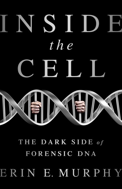
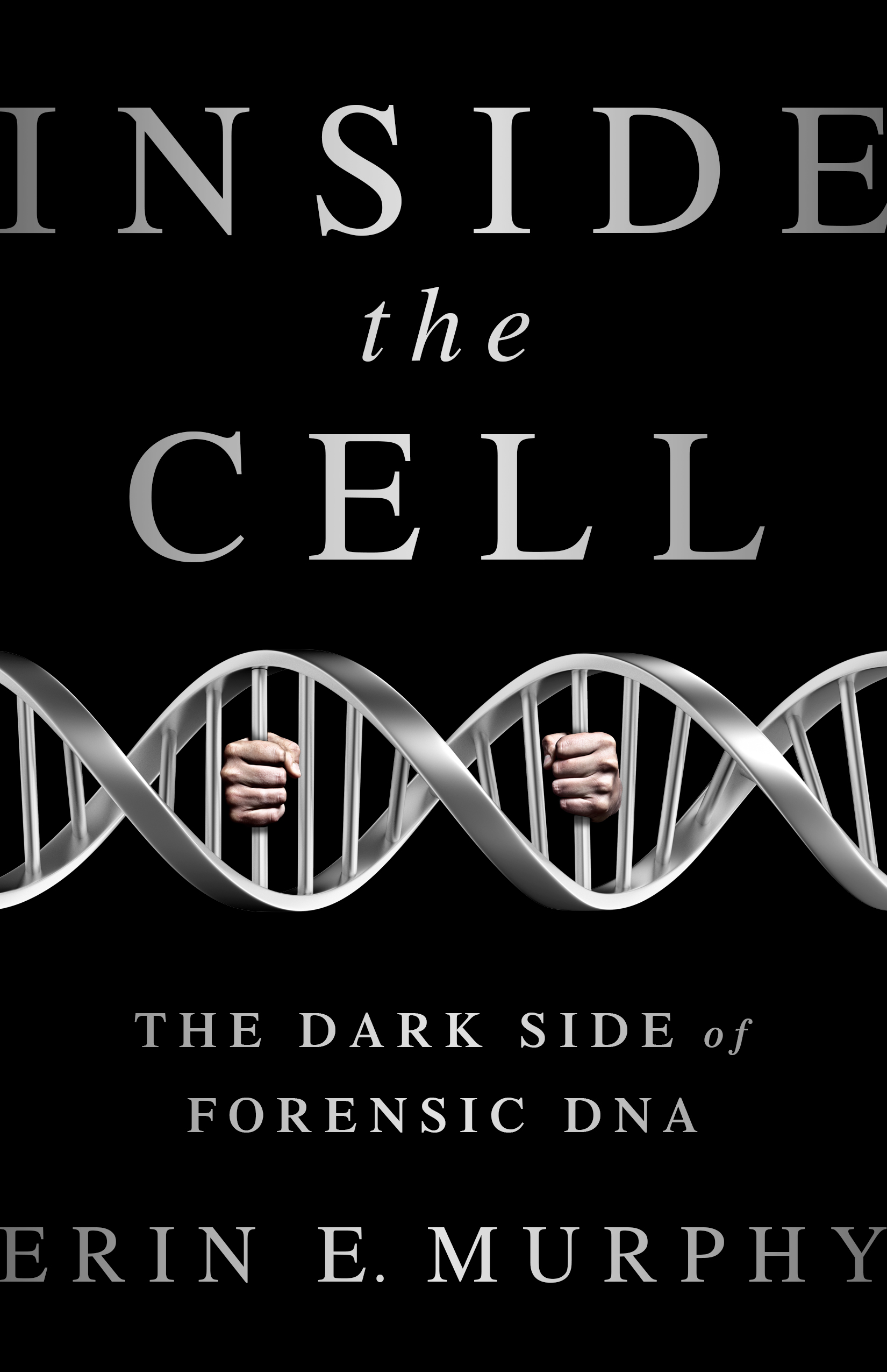
Inside the Cell

The Dark Side of Forensic DNA

Contributors

By Erin E. Murphy

Formats and Prices

Price

$14.99

Price

$19.99 CAD

Format

Format:

  1. ebook $14.99 $19.99 CAD
  2. Hardcover $27.99 $34.99 CAD

This item is a preorder. Your payment method will be charged immediately, and the product is expected to ship on or around October 6, 2015. This date is subject to change due to shipping delays beyond our control.

Josiah Sutton was convicted of rape. He was five inches shorter and 65 pounds lighter than the suspect described by the victim, but at trial a lab analyst testified that his DNA was found at the crime scene. His case looked like many others — arrest, swab, match, conviction. But there was just one problem — Sutton was innocent.

We think of DNA forensics as an infallible science that catches the bad guys and exonerates the innocent. But when the science goes rogue, it can lead to a gross miscarriage of justice. Erin Murphy exposes the dark side of forensic DNA testing: crime labs that receive little oversight and produce inconsistent results; prosecutors who push to test smaller and poorer-quality samples, inviting error and bias; law-enforcement officers who compile massive, unregulated, and racially skewed DNA databases; and industry lobbyists who push policies of “stop and spit.”

DNA testing is rightly seen as a transformative technological breakthrough, but we should be wary of placing such a powerful weapon in the hands of the same broken criminal justice system that has produced mass incarceration, privileged government interests over personal privacy, and all too often enforced the law in a biased or unjust manner. Inside the Cell exposes the truth about forensic DNA, and shows us what it will take to harness the power of genetic identification in service of accuracy and fairness.

Excerpt

PART IPART I

When a Match Isn’t a Match: How DNA Testing Goes WrongWhen a Match Isn’t a Match: How DNA Testing Goes Wrong


CHAPTER 1CHAPTER 1

The Basics: DNA Typing for DummiesThe Basics: DNA Typing for Dummies

DNA TYPING HAS resulted in the exoneration of hundreds of wrongfully convicted people, and the conviction of innumerable guilty ones. It seems like every day, the media reports a verdict overturned after new evidence exculpates the accused, or recounts the spellbinding tale of the use of science to net an elusive killer. But the stories rarely say how exactly the DNA tests helped identify the guilty or free the innocent. It would be easy to believe that DNA analysis works like a home pregnancy test—simply swab a suspect’s cheek and then read the result: guilty or not guilty.

But DNA testing is not that simple. If it were, then sending the same DNA samples to different analysts would always yield identical results. Yet studies have shown that is not always the case. For instance, in one 2011 study of subjectivity in DNA interpretation,1 researchers sent the file from an actual case that occurred in Georgia to seventeen different DNA analysts, all experienced caseworkers at an accredited government lab in the United States. The sample derived from an alleged gang rape. Given the charge, it could be assumed that the DNA samples would include at least the victim’s DNA, but it was unclear how many additional contributors might be found.

The file included the typed DNA profile of the victim along with those of three identified suspects. The analysts were asked to draw one of three conclusions for each suspect: excluded as a possible contributor to the crime scene sample, “cannot be excluded” as a possible contributor (that is, a possible participant), or “inconclusive.” One suspect in particular was considered critical: he claimed innocence, but another suspect who had confessed to the crime implicated him in exchange for more lenient treatment in his own case.

Instead of a uniform response, there was marked variation among the analysts’ findings. One examiner found that the suspect could not be excluded, and therefore his genetic material could plausibly be part of the DNA evidence. Twelve reached the opposite conclusion and excluded the suspect from having contributed to the sample. Four found the evidence inconclusive. The Georgia analyst in the actual case agreed with the lone examiner in the study who found the suspect a possible contributor—and this evidence was used to convict the real-life suspect of the crime.

Although this study was the first of its kind to be published, it simply crystallized what had long been known: interpretation of DNA from crime scenes can be incredibly complex. As Peter Gill, one of the world’s leaders in forensic science, once said, “If you show 10 colleagues a mixture, you will probably end up with 10 different answers.”2 Why were the expert’s conclusions all over the map? How is it that DNA testing allows doctors to confidently predict fetal traits, yet when DNA is studied in connection with a criminal case, even experienced, trained analysts may disagree about something as basic as who contributed to a sample?

There are five key differences between DNA science practiced in the criminal justice system—forensic DNA testing—and DNA science practiced in the medical or research context:

       1.  Forensic DNA testing involves a kind of shortcut DNA test, not the sequencing tests more typically performed in the research context.

       2.  The quantity of DNA recovered from a crime scene limits forensic analysts to what is available, whereas clinicians or researchers can take optimal quantities of samples and redraw a sample or retest if anything goes wrong.

       3.  The quality of DNA samples taken from a crime scene is often much poorer than that taken in a clinical environment or from a known individual; crime scene DNA may have degraded due to environmental exposure or contain a mix of biological material from an unknown number of people.

       4.  Forensic analysts seek to answer a question that is usually of no import to medical or clinical researchers. Namely, who is the source of this crime scene sample and how confident can we be in that attribution?

       5.  Clinical and medical researchers perform their work openly, and their results are subjected to layers of critical review before they impact individual lives. In contrast, forensic DNA testing endures far less oversight, and the DNA databases used to match suspects to crime scenes remain wholly sheltered by the government from any external critical review.

In sum, forensic DNA typing departs radically from DNA testing in the medical, clinical, or academic context. It can be a far more complex and nuanced operation than might appear at first glance. And many of these nuances and complexities are unavoidable; they arise as a matter of course in an ordinary criminal case, not just as the product of lack of training or analyst error. To truly appreciate the nature of interpretative subjectivity, one must first possess a basic understanding of the fundamentals of forensic DNA typing.

DNA TYPING: THE BASICS

Although the exact number of cells in the human body is unknown, one estimate places the number at 37.2 trillion, composed of more than two hundred different cell types.3 There are blood cells, skin cells, sperm cells, and saliva cells, among others. People shed those cells constantly. Every two minutes, in fact, we shed almost enough skin cells to cover an entire football field.4 A typical human fingerprint, barely visible to the naked eye, contains roughly one hundred cells.5

Inside nearly every one of those cells lies a nucleus,6 and inside that are twenty-three sets of paired chromosomes that form the human genome. One chromosome in each pair is inherited from one’s biological mother, and the other from one’s biological father. The sex chromosomes that make up the final pair determine whether a person is genetically female (XX) or male (XY).

Unfurled, these DNA strands stretch roughly three feet long in the form of the characteristic double helix structure that won James Watson, Francis Crick, and Maurice Wilkins the Nobel Prize in 1962.7 You can imagine the DNA strand as a twisting ladder, in which each rung is a step joining two base pairs. There are only four possible bases—abbreviated A, T, C, and G—and they always pair off in the same way: A joining with T, and C joining with G. In the 1990s, the Human Genome Project started a global race to assemble the human genome: participating scientists tested one person’s DNA sample to figure out every single letter along each side of that ladder. There are 3.2 billion such rungs, so it is easy to see why the project to discover, or “sequence,” them all did not finish until 2003.8

At first, researchers assumed that the human genome would differ dramatically from that of other organisms. But as scientists sequenced more human and nonhuman genomes, it turned out that huge chunks of the human genome were identical to those of cows (85 percent), dogs (84 percent), and chickens (65 percent). Indeed, we share half of our genes with the common fruit fly, and a quarter of them with a grain of rice or a wine grape!9 Given how much we have in common with plants and animals, it is perhaps not surprising that the variation in the human genome is incredibly slight—roughly 0.1 percent.10 But although it is slight when expressed as a percentage, it still is significant in absolute numbers. That percentage encompasses roughly 3 million base pairs of difference.

It would be easy to assume that because so little of the genome varies from person to person, the parts that do differ must all be critical in determining any one person’s makeup. In other words, you might think that this 0.1 percent is packed wall to wall with genes. Genes, as most people know, are the identifiable stretches of the genome that we know have a clear function in the human body. Typically thousands of base pairs in length, genes determine characteristics as superficial as eye color or as profound as an increase in susceptibility to breast cancer.11

But the 0.1 percent is not full of genes. It is estimated that there are only about 25,000 genes. The purpose of the base pairs contained within the remaining areas of variation is currently unknown; indeed, huge stretches of the genome do not seem to play any role in the actual function or appearance of the body, even though they differ from person to person. In some ways, this finding nicely dovetails with another recent realization: that even parts of the genome that are identical may nonetheless have different manifestations when actually put to work.

Researchers interested in medicine and human biology typically study the working—or coding—parts of the genome, for obvious reasons. Academic and medical researchers are interested in finding clues that might prevent or treat disease or disabling conditions, or even just help understand why some people lose their hair while others do not. That means that, for the most part, these researchers also engage in sequencing of genes. They want to learn the specific letters that make up the ladder rungs of the double helix, along with their order, because the letters are like the instruction manual to the rest of the body. If scientists can uncover how to read the instructions, they will be able to rewrite them when they go awry.

Forensic DNA testing, in contrast, deliberately ignores the genes, analyzing only those regions of the genome that vary for no discernible reason. That is, forensic DNA methods focus on the noncoding regions of the genome—the parts that do not send any orders. That choice is made intentionally, most notably to protect privacy and ensure accuracy. Looking at the noncoding parts also gives forensic scientists greater confidence that each place they look is disconnected from the other places studied. Think about it in terms of coding genes: we know from experience that blue eyes, blond hair, and light skin tone tend to go together more than do blue eyes, brown hair, and dark skin tone. That means that there may be genetic connections between those traits—such that having blond hair makes it more likely that one will also have blue eyes. If they are connected, they are not independent of one another—and that diminishes their value in distinguishing one person from the next. To maximize the use of genetic information to identify people, we want as many disconnected data points as possible; otherwise the value of each point is compromised by its connection to another piece of information that predetermines its result.

The other important way in which forensic DNA typing differs from medical or academic research is that forensic DNA typing usually ignores sequence variation. That is, it does not differentiate among people by typing the base pair sequences. Instead, forensic DNA typing studies a different kind of variation—specifically, length variations.

FORENSIC DNA TESTS: A PRIMER

Since forensic DNA methods first emerged in the 1980s, several techniques have been used. Most common today is an approach that looks at short, tandem, repeat sections of the genome—known as STR typing. To understand STR typing, some basic vocabulary is essential. First, recall that the word gene denotes a section of the genome that has some purpose or function—each gene regulates the body in some way. A more general term for these sections is the Latin word for “place”—locus, or loci in the plural. The word locus is the preferred way of referring to identifiable chunks of the genome that have no purpose but are distinguishable for other reasons (most pertinently, because they vary from person to person). Forensic DNA typing typically uses thirteen loci as its core set of identifiers, although that number will soon be raised.12

Here is the tough part. At each of these loci, known sequences repeat in different combinations in different people. These known sequences are short—typically around four base pairs—and they are the same in every person. The only difference is that they repeat a different number of times in each individual in predictable increments.13

The more the sequence repeats, the longer that fragment of DNA. Thus, by measuring the length of the fragment, an analyst can identify the genetic “signature” of a person at that locus. This is done by analyzing two repeat lengths, one from each biological parent. Some loci have as few as eight commonly observed patterns of repeats (for example, the sequence repeats anywhere from ten to seventeen times), whereas others have as many as twenty-seven. By counting the number of repeats present at each pair of the thirteen loci, an analyst can thus obtain twenty-six discrete measurements, or variations, for an individual.14

These variations are called alleles. So, if a suspect has a “5, 8 allele pattern at the D3 locus,” this means that at the locus D3, the suspect has five repeats of a known sequence on one of the chromosomes, and eight repeats on its partner chromosome. When all the alleles of the selected loci are compiled, the result is the individual’s genetic profile—a composite of traits that identify that person. Because each of the thirteen loci has anywhere from eight to twenty-seven different allele possibilities, and each person has two alleles at each locus,15 the genetic “signature” becomes highly discriminating. Only identical twins share an entire genome, and so examining a sufficient number of snippets of the genetic strand typically distinguishes one person from another.

FORENSIC DNA TESTING

How is DNA obtained and tested forensically? It starts, of course, with a sample. For controlled samples, such as those from known persons, the most common approach today is what is known as a buccal swab. This is simply a painless scraping of the inside of a person’s cheek to collect skin cells, using a cotton swab–like sampling device. Uncontrolled samples, such as crime scene evidence, are usually collected in either of two forms: a swab or a cutting. Swabs are simply the rubbing of those sampling devices against a surface (say, of a light switch or bloodstain), whereas a cutting is a piece of an item likely to contain biological material (for instance, fabric or upholstery). Often referred to in shorthand as stains, these swabs or cuttings most commonly hold biological material such as blood, skin (epithelial cells), sperm, or saliva—but any human cell with a nucleus can yield a result (even sweat, tears, etc.). That means that a wide array of objects can yield DNA results: predictable things, such as guns, bloody clothing, or an intimate swab containing ejaculate; but also unexpected sources, such as a sweat-stained hat brim, a half-eaten burger, a used facial tissue, or a disposable razor.

In the early days of forensic DNA typing, an analyst needed a significant amount of biological material to conduct a test likely to yield a useable result. For instance, if you can picture a dime, the forensic sample would have needed to take up about half of the dime’s surface. Today, however, much less is needed; a sample the size of just one of the digits in the minuscule year printed on a dime can reliably produce a profile. Techniques for maximizing the recovery of genetic material from crime scene stains continue to evolve, and some newer methods even extract genetic material from as little as a dozen or fewer cells.

Once an analyst has a crime scene stain in hand, the next step is to figure out what kind of sample it is and whether it contains human DNA. In a process called extraction, the analyst separates the DNA from all the other cellular materials that make up the sample. The nucleus of the cell is broken up and all the extraneous bits washed away (there are several different methods for this, most involving chemical washes), exposing the DNA left behind. This reduces the stain to a DNA sample. Because extraction is highly sensitive, it has been described as “probably the moment where the DNA sample is more susceptible to contamination in the laboratory than at any other time in the forensic DNA process.”16 Due to this risk of contamination and the difficulty of uncovering it after the fact, it is crucial that labs take care not to unintentionally introduce foreign material to the sample. Good labs will use different stations for processing evidence samples (for example, the murder weapon) and reference samples (such as the suspected killer’s buccal swab), and sequence and separate the testing process so that the entire area may be thoroughly cleaned, and so that at worst any problems arise in a sample of known origin as opposed to a crime scene sample.

Special methods of extraction help produce results in difficult cases. One method was proposed in 1985, by Peter Gill.17 Known as differential extraction, this process solves a problem common in rape cases: intimate swabs from sexual assaults often contain a mixture of material. For instance, a vaginal swab may contain traces of the attacker’s ejaculate, but may be overwhelmed by cells from the victim. Differential extraction allows the analyst to segregate the sperm portion of the sample from the skin cell portion of the sample, so that the analyst can distinctly see the male portion of the mixed sample. If there is no sperm in the sample, another way of separately examining the male fraction is to look for markers specific to the Y chromosome.

The next step in the testing process is known as quantification. Different biological sources carry different amounts of DNA—liquid blood or semen has a large quantity (roughly 20,000 to 40,000 nanograms [ng]), whereas urine or bone contain much less (roughly 1 to 20 ng).18 Before testing, the analyst must ensure that the sample contains an appropriate quantity of DNA. The amount matters because DNA testing machines are sensitive: too much or too little DNA will compromise the results.

Next comes the core of forensic DNA testing—two processes known as amplification and capillary electrophoresis. Amplification accomplishes two things: first, it reduces the 3.2 billion base pair genome down to the particular fragments that the forensic analyst is interested in, and second, it copies just those segments so that they may be examined more closely. Some people liken this process to genetic Xeroxing, but it actually trims down the sample first, before building it up again in as section-specific replicates.

To illustrate, imagine you went to watch your friend run in a marathon. You want to make sure you see her when she passes by, so you can meet her at the end of the route. If your friend is going to be running on her own, it might be easy to miss her in the crowd. If you turn away for a minute, or if your attention is captivated by someone else along the route, she might jog by without your noticing. But if your friend plans to run with a large team dressed identically, she would be almost impossible to miss. Even if you did not see her specifically, you would know for certain when she passed because her group would be hard to overlook.

DNA testing works along a similar principle: the pertinent fragments of DNA are so tiny that measuring them correctly, if you looked at just one, might be difficult. Indeed, this problem would be particularly vexing in forensic cases, because not only are the fragments small, but the number of available fragments may be few. With amplification, however, a small amount of DNA can be trimmed down to the relevant bits, and then replicated to millions or even billions of copies that make studying those little fragments possible.

This genetic photocopying technique, known as polymerase chain reaction, or PCR for short, so revolutionized molecular biology that its inventor received the Nobel Prize.19 In forensic typing, commercial kits are available that contain primers, tiny chemical scissors that help cut the whole genomic strand into just those tiny fragments of interest to the analyst. The PCR process then applies alternating cycles of heating and cooling (typically 28 to 32 cycles20) that copy those fragments. During the amplification, the samples are also tagged with fluorescent labels in different dye colors to separately identify each part.

Amplification thus achieves several goals: it cuts the genomic strand down to just the parts the analyst is interested in measuring, copies those parts so that there are more of them, and tags the genetic material so that it can be measured. Scientists continue to improve upon amplification techniques, such as improving the ability to amplify multiple spots on the genome at one time. Known as multiplexing, these methods permit a scientist to use a small amount of genetic material to create a large amount of selectively copied regions all in one round of PCR. However, multiplex processes also introduce added sensitivities, as the ideal conditions for amplifying one region of the genome may not perfectly mirror the conditions needed for amplifying other regions.

Other problems can arise during PCR that result in the loss of genetic material. Just as the quality of an image declines as you make photocopies from photocopies rather than from the originals, so, too, can the quality of genetic copies lose their integrity. Pieces of the DNA strand may slip, leaving genetic replicates close in size to the original material but slightly shorter. Or a mutation in the parts of the genome adjacent to the portion of interest can cause the primers—those genetic scissors—not to work, leading to a failure to amplify that region. The primers may likewise fail if some non-DNA material manages to infect the sample and inhibit their proper functioning.

Contamination at this stage is also a real concern; the addition of even the tiniest amount of extrinsic DNA to the sample before amplification will result in the extra DNA being copied many times over, making it seem like part of the original sample. In fact, the risk of inadvertent contamination is so great that most labs maintain databases with the genetic profiles of staff members and even crime scene investigators, so that accidental contamination with their material will be immediately apparent.

Once the DNA sample has been cut up into the relevant pieces and those chunks have been reproduced, the next step is to measure the size of the resulting DNA fragments. This is typically done through a second process, capillary electrophoresis. Electrophoresis accomplishes two main tasks. First, it separates out clusters of like material from the soup of DNA fragments that was created in PCR. Separation is a delicate process because large multiplexing systems can process so many fragments at the same time. It is akin to throwing a large family’s laundry into one basket—it not only makes it easy to lose a sock here and there, but also may lead to mistakenly putting Big Sister’s leggings in Little Sister’s drawer. These dangers are particularly acute given that some alleles differ very minimally.21

The second major task of electrophoresis is to measure the fragments to determine the allele that is present. This measurement is accomplished by effectively entering the fragments in a race. Again visualize a marathon with hundreds of entrants. Some runners are small and sprightly and will finish quickly; others are larger and move more slowly. If you are not wearing a watch, but want to find out how long it takes for certain runners to cross the finish line, you might look for pace setters. Those runners, planted in the race to run at a specific speed, offer a point of comparison for determining the rate of the unknown entrants. Because you know that a certain pace setter will finish in three hours, you know that the unknown runner that finishes next to the pace setter also must have run the course in three hours.

Electrophoresis operates much the same way. To get the DNA fragments started, a negative electric current is applied to the sample. That current causes the DNA to move toward a positive current at the other end of the capillary, or tube. Along with the material that is being tested, the analyst runs a sample called an allelic ladder, which functions as the pace setter. As each of the strands get to the positive side, a detection window makes a note of the point at which the fragment crossed and the color of the fluorescent tag that was attached during the amplification stage. The fragments are then measured against the allelic ladder, which contains known size markers (the pace setters). To make sure the process runs smoothly, the analyst also runs several controls, including a “negative control” that ensures that there are no contaminants in the capillary, a “positive control” that contains DNA with a known profile to ensure that the test yields accurate results, and an “amplification blank” to ensure that no extraneous material compromised the amplification stage.

The next chapter and the Appendix both provide more detail about the capillary electrophoresis process, and the kinds of challenges an analyst may encounter when testing a sample. But this overview gives sufficient foundation in the basic testing technique. The machines most commonly in use today take about thirty minutes per run, but more rapid machines capable of returning results within 90 minutes, measured from swab-in to the profile output, are increasingly available.22

DATABASES

Genre:

  • “A specialized work that will appeal to attorneys, investigators, crime writers, and others on the frontiers of forensic DNA laws and technologies.” –Kirkus Reviews

    “This brilliant, accessible, and extremely important book by one of our foremost scholars has a timely message: Without disputing its transformative impact, Murphy argues that DNA testing is not a panacea for the criminal justice system or forensic scientists. Murphy provides a balanced, thoughtful, and essential roadmap to a more democratic future. A must-read.” —Barry Scheck and Peter Neufeld, cofounders, the Innocence Project

On Sale
Oct 6, 2015
Page Count
400 pages
Publisher
Bold Type Books
ISBN-13
9781568584706

Erin E. Murphy

About the Author

Erin Murphy is a professor at NYU School of Law and an expert in DNA forensics. She is a graduate of Harvard Law School. In addition to scholarly journals, her writing has appeared in Scientific American, New York Times, USA Today, Slate, San Francisco Chronicle, and Huffington Post. Follow her on Twitter @ErinMurphysLaw.

Learn more about this author